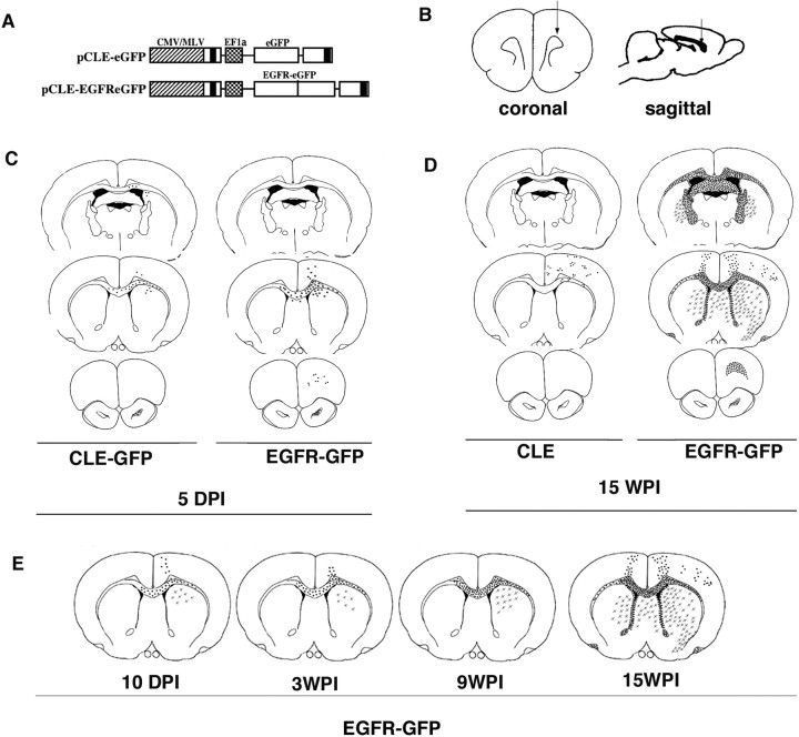
Figure 1.
EGFR-GFP constitutive expression enhances migration of infected cells. A, B, White matter glial progenitor cells were infected with either the control virus pCLE-eGFP or the pCLE-EGFReGFP (A) by unilateral viral injection to the rostral white matter (B) at P3. C, At 5 dpi, EGFR-GFP+ cells had migrated farther than control, CLE-GFP-infected cells, both rostrally and contralaterally through corpus callosum (middle panels). At 15 wpi, CLE-GFP cells have migrated through white matter and cortex, mainly on the ipsilateral side of the injection. D, In contrast, EGFR-GFP+ cells widely infiltrated the brain both ipsilaterally and contralaterally, mostly along white matter tracts (corpus callosum, fimbria-fornix), reaching rostral white matter and olfactory bulb (bottom right). EGFR-GFP+ cells invaded gray matter, but preferentially the ventral structures: caudate–putamen and striatum. E, Analysis of the EGFR-GFP+ cell distribution at different time points (10 dpi, 3 wpi, 9 wpi, and 15 wpi) showed the continuous increase in the distance that cells had migrated from the injection site.

