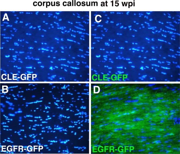
Figure 6.
Increased cell number in corpus callosum in EGFR-GFP-infected animals at 15 wpi is a consequence of the increase in the number of GFP+ cells only. Cell nuclei were stained with Hoescht dye (blue) (A–D), and GFP+ cells fluoresce green (C, D). At 15 wpi in CLE-GFP-infected animals (C), there were no GFP+ cells in the center of the corpus callosum. In contrast, in EGFR-GFP-infected animals GFP+ cells had widely infiltrated the callosum in large numbers (D). The total cell number in the corpus callosum was more than twice that of control, and the excess cell number was made up entirely of GFP+ cells (see Results).

