Figure 7.
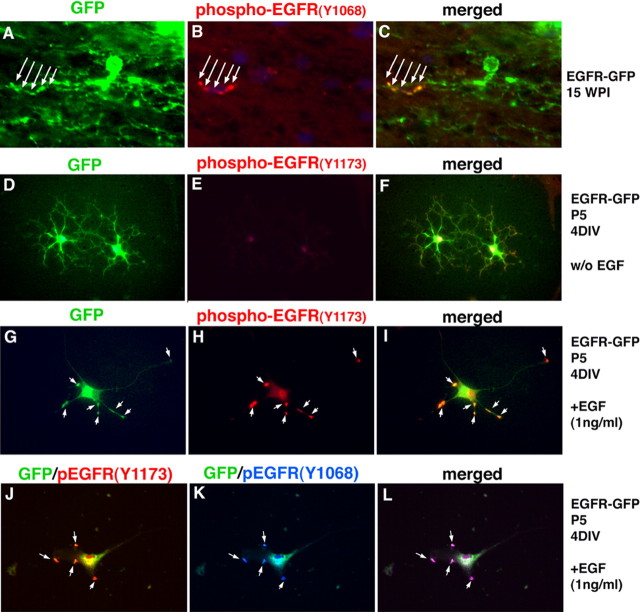
EGFR-GFP-infected cells signal actively in vivo an in vitro. A–C, GFP was found in specific foci on the processes of EGFR-GFP-infected cells in white matter (A), consistent with receptor accumulation. B, C, Some of the GFP+ accumulations were also positive for anti-phospho-EGFR (Y-1068) antibody. D–I, Glial progenitors infected with EGFR-GFP and grown in the basal media for 4 d showed basal levels of EGFR phosphorylation (Y1173; D–F), but in the presence of EGF (1 ng/ml; G–I), the level of phosphorylation increased dramatically. GFP signal on the membrane overlaps with the positive immunostaining for P-EGFR (arrows). J–L, Double immunostaining for two phosphorylated tyrosine residues (Y1068 and Y1173) (arrows).

