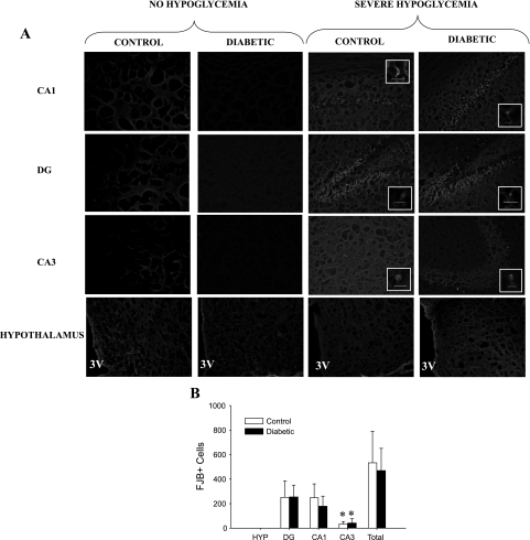
Fig. 5.
Hippocampal and hypothalamic neuronal damage. A: FJB+ staining in the CA1, DG, and CA3 regions of the hippocampus and the hypothalamus viewed at ×100 magnification. A, insets: FJB+ cells viewed at ×400 magnification, with the bars indicating 100 microns. The pictures of the hypothalamus (HYP) were taken lateral to the 3rd ventricle (3V). The 4 groups shown are nondiabetic control and STZ-diabetic rats that did not undergo hypoglycemia, nondiabetic control rats that were subjected to severe hypoglycemia, and STZ-diabetic rats that were subjected to severe hypoglycemia. B: in response to severe hypoglycemia, both control (n = 5) and diabetic (n = 8) rats demonstrated no neuronal damage in the hypothalamus, but hippocampal neuronal damage was most evident in the DG and CA1 regions, with significantly less damage in the CA3 region. *P < 0.05.

