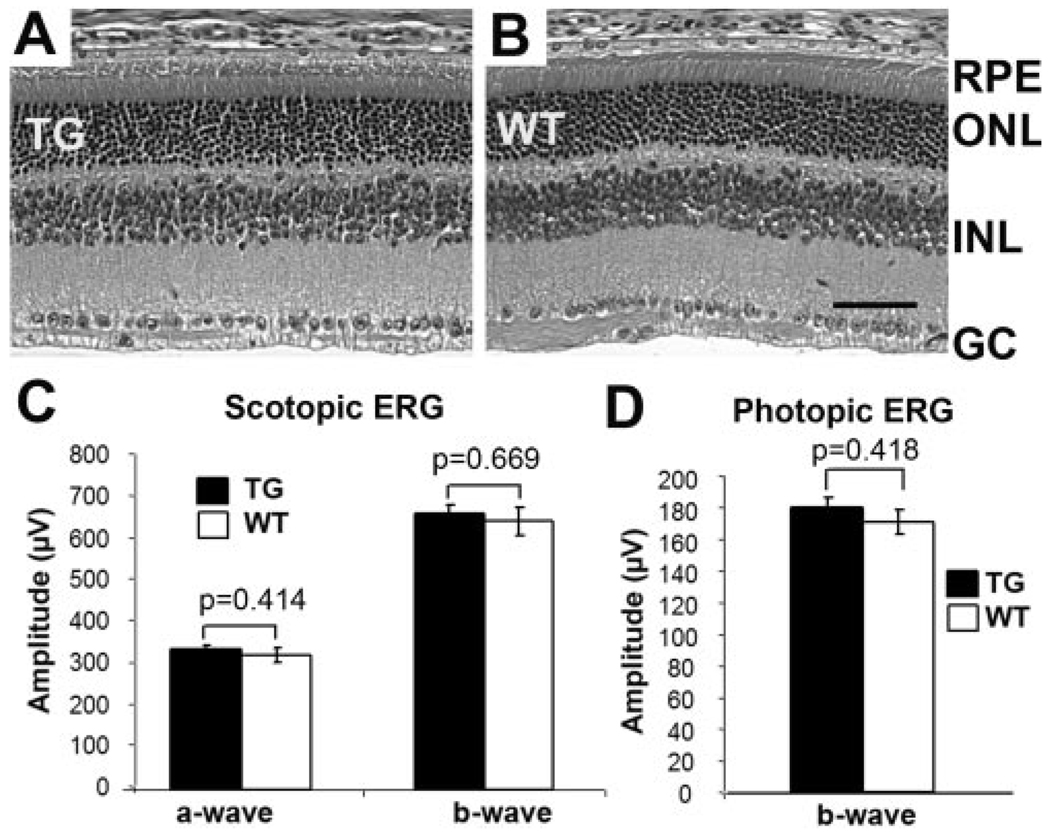
FIGURE 5.
Retinal morphology and function of 10-month-old inducible RPE-specific Cre mice. (A, B) Representative H&E-stained retinal sections of transgenic (TG) and wild-type (WT) mice. RPE, retinal pigmented epithelium; ONL, outer nuclear layer; INL, inner nuclear layer; GC, ganglion cells. Scale bar, 40 µm. (C, D) Scotopic and photopic ERGs. At least eight mice were included in each group. No significant difference in retinal morphology and function was detected in the inducible RPE-specific Cre mice.

