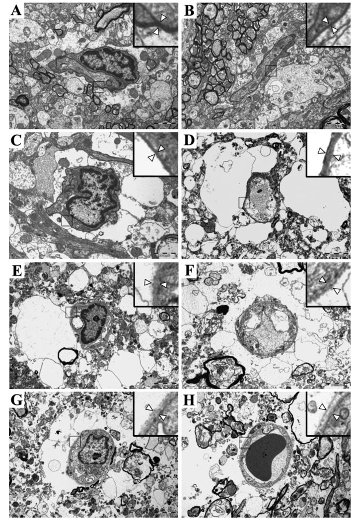
Fig. 4.
Representative electron microscopic photographs. A: Sham control. Almost all surface areas of the microvessel are covered by astrocyte endfeet. B: One hour after middle cerebral artery occlusion (MCAO). C: Four hours after MCAO. Swelling of the astrocytes and focal detachment of the astrocyte endfeet from the basement membrane (BM) are seen. D: Eight hours after MCAO. A marked astrocyte swelling and a further decrease in the intact portion of the contacts are demonstrated. E: Twelve hours after MCAO. The proportion of the intact BM–astrocyte contacts is markedly decreased, and the BM is damaged. F: Sixteen hours after MCAO. Further degradation of the BM and ruptured astrocytes is evident. G: Twenty hours after MCAO. There is excessive accumulation of water around the microvessels. H: Forty-eight hours after MCAO. The astrocyte endfeet are no longer visible, and the BM is very faint. Arrowheads in the inset (magnified view) indicate the BM. Original magnification ×8,000.

