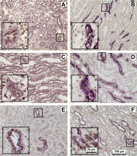
Fig. 3.
Immunolocalization of HuR in kidney sections of sham-operated rats. Using double label immunohistochemistry, HuR (shown in brown) nuclear localization was observed in all renal tubules identified by segment-specific markers (shown in purple). The proximal tubules and the thin descending limbs of Henle were identified as immunoreactive cells for aquaporin-1 (AQ1) in the cortex (A) and medulla (B), respectively. The thin ascending limb of Henle (C) was distinguished in the medulla by the presence of chloride channel-K1 (CLC-K1). The thick ascending limb was identified as Tamm-Horsfall glycoprotein (THP)-positive cells in the medulla (D). The thiazide-sensitive Na+-Cl+ transporter (TSC) was used as a marker of distal convoluted tubules in the cortex (E). Aquaporin-2 (AQ2) antibodies were used to visualize collecting tubules (F). Insets: marker-positive cells in higher magnification.

