Fig. 3.
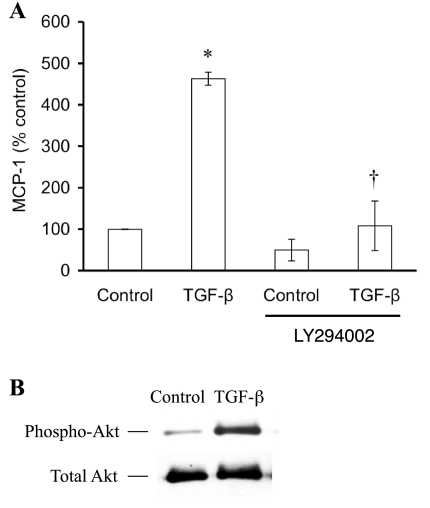
Phosphatidylinositol 3-kinase (PI3K) mediates TGF-β1-stimulated MCP-1. A: a specific inhibitor of PI3K, LY294002 (25 μM), completely inhibited TGF-β-stimulated MCP-1 production by cultured podocytes (n = 3). The modest decrease in MCP-1 due to LY294002 alone was not significantly different from control. *P < 0.05 vs. control. †P < 0.05 vs. TGF-β. B: TGF-β1 treatment of podocytes activated the PI3K pathway, evident in the increased amount of phospho-Akt compared with the constant level of total Akt.

