Fig. 4.
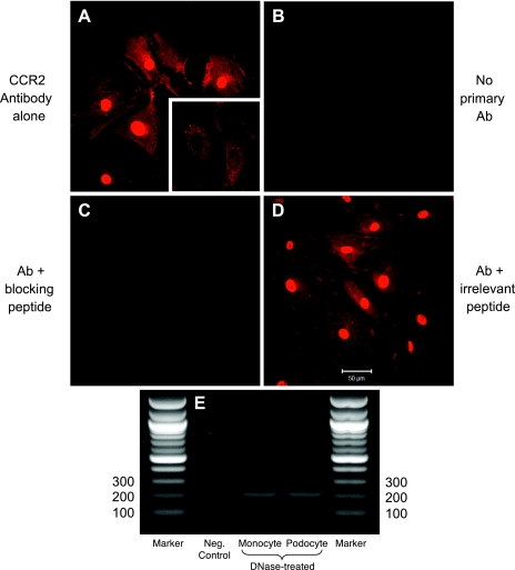
Cysteine-cysteine chemokine receptor 2 (CCR2) protein and mRNA in podocytes. A: CCR2 staining is evident in podocytes as a red signal. The intense nuclear signal abates when the cells are not permeabilized before staining (inset). B: no fluorescence is detected when the primary antibody is omitted. C: staining is competitively obliterated by a blocking peptide, indicating the specificity of the primary antibody for CCR2. D: staining is not affected, however, by an irrelevant blocking peptide (in this case, VEGFR-1 antigen). Magnification: ×400. E: RT-PCR confirms the expression of CCR2 mRNA in podocytes (200-bp band). RT-PCR performed on monocyte RNA, a positive control, shows a CCR2 band of identical size. The negative control, water, showed no RT-PCR band.

