Fig. 7.
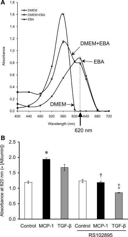
MCP-1 increases podocyte permeability to albumin. A: Evans blue-labeled albumin (EBA) can be quantified by the absorbance characteristics of the Evans blue dye, which absorbs light most strongly at 620 nm. In our tests, the A620 measurement is oblivious to DMEM but is quite sensitive to the mixture of DMEM+EBA. B: in a Transwell setup to assay the cellular permeability to EBA, performed after the transepithelial electrical resistance had plateaued at 114.5 ± 24.6 Ω·cm2, significantly more EBA had diffused from the lower into the upper chamber across the podocyte monolayer at 24 h as a result of MCP-1 treatment (50 ng/ml) vs. control. TGF-β (2 ng/ml) had a similar effect on EBA transit, although not statistically significant. The permeability to albumin induced by MCP-1 or TGF-β was returned to control levels by concurrent treatment with RS102895 (6 μM), an inhibitor of CCR2 signaling (n = 4). *P < 0.05 vs. control. †P < 0.05 vs. MCP-1. ‡P < 0.05 vs. TGF-β.

