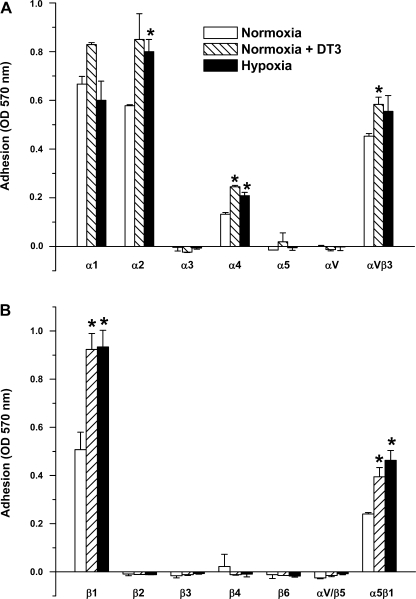
Fig. 2.
α-Integrin (A)- and β-integrin (B)-mediated binding profile of FPASMC exposed to normoxia, hypoxia, or normoxia in the presence of PKG inhibitor DT-3 (3 μM) for 24 h, harvested, and incubated for 1 h at 37°C in wells coated with anti-α- or anti-β-integrin monoclonal antibodies. Values are means ± SE (n = 3). *Significantly different from respective values observed in control (P < 0.05).

