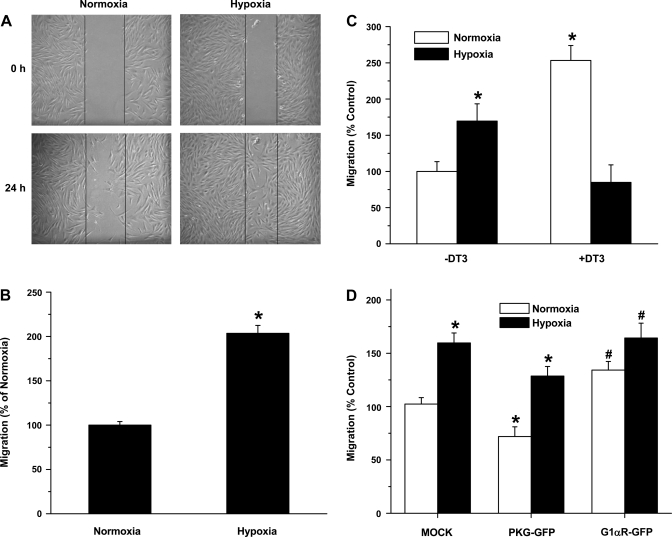
Fig. 5.
Downregulation of PKG in hypoxia or inhibition of PKG in normoxia with a dominant- negative PKG construct (G1αR-GFP) or PKG1 inhibitor DT-3 (3 μM) leads to increased FPASMC migration. A: hypoxia-induced increase in migration of FPASMC by wound healing model. Scratch on cell monolayer (0 h) and migration at 24-h normoxia and hypoxia. B: migration was assessed at 24 h by counting the number of cells that migrated across the scratch, normalized to scratch area and presented as %, with normoxia set at 100%. Values are means ± SE (n = 4). C: Boyden chamber migration assay of FPASMC treated with normoxia, hypoxia, or normoxia in the presence or absence of PKG1 inhibitor (DT3, 3 μM). Migration is presented as %, with normoxia (−DT3) set at 100%. Values are means ± SE (n = 3). *Significantly different from normoxia (−DT3) value (P < 0.05). D: Boyden chamber migration assay of FPASMC transfected with vector alone (pEGFP-N1, mock), PKG-GFP, or PKG 1αR-GFP (dominant negative) constructs. Migration is presented as %, with vector set at 100%. Values are means ± SE (n = 3). *Significantly different from respective normoxia values (P < 0.05); #significantly different from mock, normoxia value (P < 0.05).

