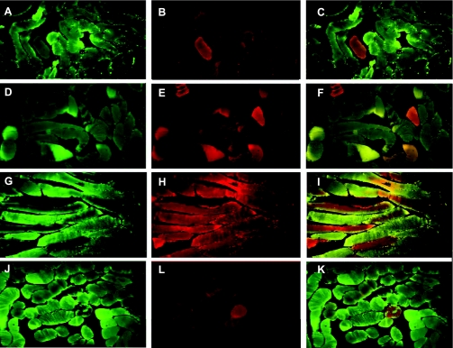
Fig. 3.
Immunofluorescence microscopy of skeletal muscle. COX-I (green fluorescence-mitochondrial encoding) and succinate dehydrogenase (SDH) subunit A (red fluorescence-nuclear encoding) were stained with specific antibodies, and the images were merged to assess colocalization. A–C: typical SDH (A) and COX-I (B) staining of control muscle and the overlay (C). D–F: typical stained muscle sections for SDH (D), COX-I (E), and the overlay (F) from the air plus V̇o2max group. G–I: muscle sections from the CO plus V̇o2max group stained for SDH (G), COX-I (H), and the overlay (I). J–K: SDH (J) and COX-I (L) staining and the overlay (K) in a sample of muscle after 5 days of CO breathing.

