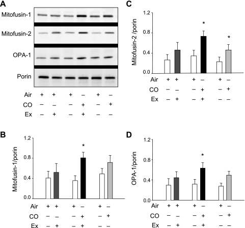
Fig. 5.
Mitochondrial fusion [mitofusin (mFn)] proteins and optic atrophy (OPA)-1 responses to inhaled CO with or without V̇o2max testing. A: Western blots of muscle tissue for mFn1, mFn2, and OPA-1 before and 5 days after inhaled CO with or without the V̇o2max test. Relative protein expression was derived by normalizing mFn1, mFn2, and OPA-1 to porin, a stable mitochondrial outer membrane protein. B: relative mFn1 expression. C: relative mFn2 expression. D: relative OPA-1 expression. Values are means ± SD (*P < 0.05 between groups).

