Fig. 3.
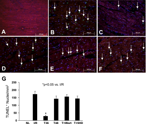
Representative photomicrographs showing terminal deoxynucleotide transferase-mediated dUTP nick end labeling (TUNEL)-positive nuclei in various treatment groups. A: normal heart tissue. B: TUNEL-positive nuclei (green, white arrow) in I/R control group. C: tadalafil-treated group at 36 h (T36). TUNEL-positive nuclei were significantly decreased in tadalafil-treated hearts compared with those in the I/R group. D: tadalafil-treated group at 48 h (T48) showing reappearance of apoptosis. E: tadalafil + Wort treatment group. F: tadalafil + 5-HD treatment group. Apoptosis is similar to I/R group (B). All nuclei were stained blue by 4,6-diamidino-2-phenylindole, and heart tissue was stained with α-sarcomeric actin (red) for cardiomyocytes. Magnification, ×400; n = 4 animals for each group. Bars = 100 μm. G: quantitative estimate values of TUNEL-positive nuclei in data are means ± SE. *P < 0.05 vs. I/R Control; n = 4 animals for each group.

