Fig. 1.
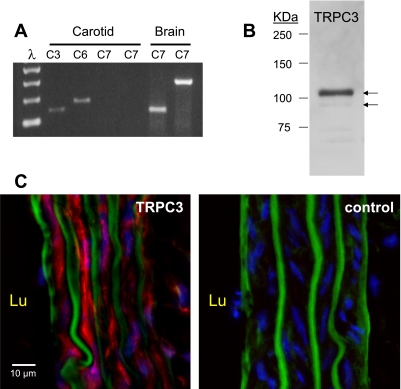
A: evaluation of transient receptor potential channel (TRPC) 3/6/7 mRNA expression in rat carotid artery (CA) by RT-PCR. PCR amplicons of predicted size were detected for TRPC3 and TRPC6, but not for TRPC7. Brain cDNA was used as a positive control for the TRPC7 primers. A 100-bp ladder is shown to the left. B: evaluation of TRPC3 protein expression in rat CA by Western blot. The arrows indicate the doublet corresponding to the glycosylated and nonglycosylated forms of the TRPC3 protein. C: immunofluorescence analysis of TRPC3 expression in CA frozen sections. TRPC3 expression (red) is present throughout the smooth muscle layers, but is not seen in the endothelial cells. The green signal reflects the elastic laminae recorded as the autofluorescence in the green channel. Cell nuclei appear in the blue channel. Lu, luminal (endothelial) side.

