Fig. 3.
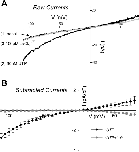
A: representative current-voltage (I-V) plots in freshly isolated rat CA smooth muscle cells (SMCs). The currents reflect the baseline current (1), the current following application of 60 μM UTP (2), and then the cumulative application of UTP and 100 μM La3+ (3). B: summary of the UTP-stimulated current (IUTP) (subtracted currents; UTP-basal) before (diamonds) and after application of La3+ (circles). Currents are normalized to cell capacitance. Administration of La3+ abolished the IUTP.

