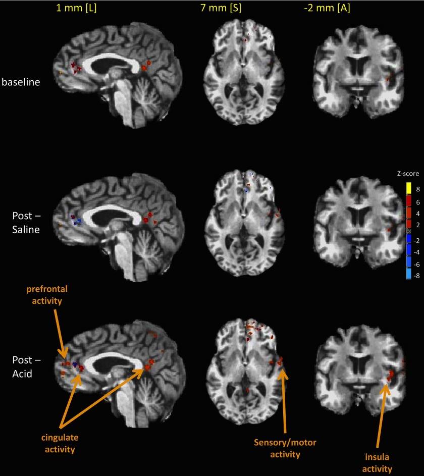
Fig. 5.
Locations of cerebral cortical swallow-related activity resulting from group analysis of all tested subjects are shown as colored areas superimposed on stereotaxic, high-resolution MR anatomical images. All data were transformed to the Talairach-Tournoux coordinate reference frame. Images are shown in the sagittal [1 mm left (L) in the stereotaxic coordinate frame], axial [7 mm superior (S)], and coronal [−2 mm anterior (A)] radiographic views. Different sagittal, axial, and coronal views are shown to illustrate all fMRI regional activity. As seen, there is greater cortical recruitment and increased signal intensities for swallow-related fMRI activity in the cingulate, sensory-motor, prefrontal, and insular region of interest after esophageal acid stimulation (postacid) compared with after saline perfusion and preinfusion (baseline).

