Figure 3.
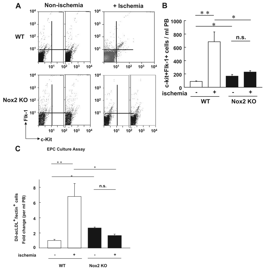
The number of circulating EPC-like cells after hindlimb ischemia is inhibited in Nox2−/− mice. A, The number of EPC-like ckit+Flk1+ cells in peripheral blood (PB) mononuclear cell fraction at 0 (+ nonischemia) and 3 days (+ ischemia) after hindlimb ischemia in WT and Nox2−/− mice, as measured by FACS analysis. B, Statistical analysis of ckit+Flk1+ positive cells in PB (n=6, *P<0.05, **P<0.01). C, EPC culture assay. Quantitative analysis of the numbers of DiI-acLDL and BS lectin double positive EPCs measured at 4 days after culture of PB-derived MNCs obtained from WT and Nox2−/− mice at 0 and 3 days after ischemia. Data are expressed as fold change over basal (the ratio on day 0 in WT mice was set to 1). (n=4 to 6, *P<0.05, **P<0.01).

