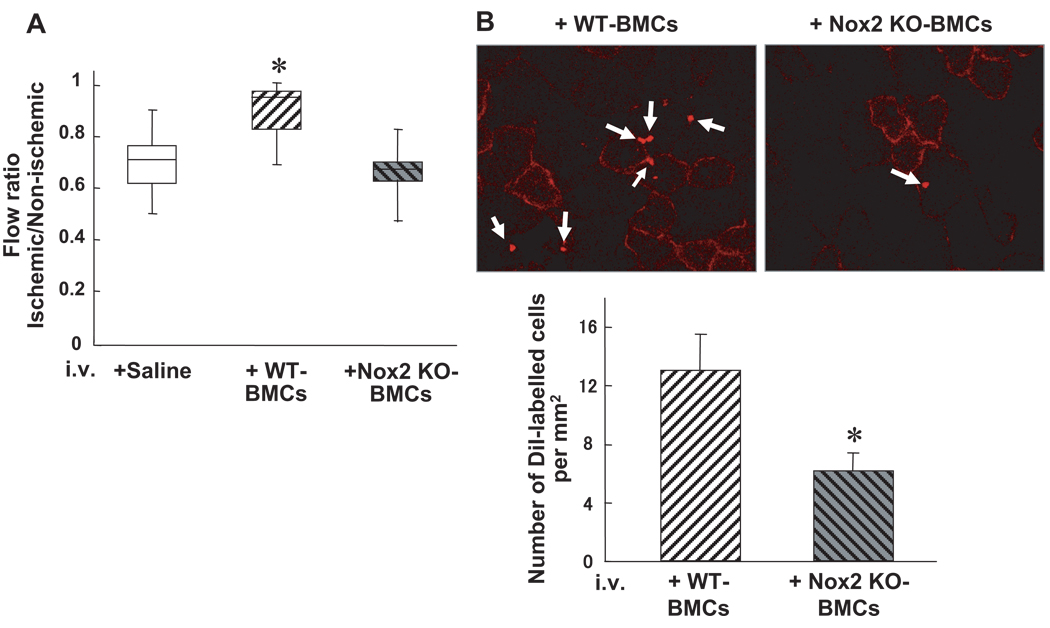
Figure 5.
Homing and neovascularization capacity are impaired in Nox2−/− BMCs in vivo. A, BMCs (3×10⁶ cells) from WT or Nox2−/− mice, or saline control, were intravenously injected into WT mice at 24 hours after femoral artery ligation, and blood flow recovery, as assessed by the ischemic/nonischemic LDBF ratio, was measured at 14 days after hindlimb ischemia. (n=4, *P<0.05 vs control). B, Upper panel, representative photographs of accumulated intravenously injected Dil-labeled WT- and Nox2−/− BMCs in ischemic adductor muscles. Lower panel, quantitative analysis of the number of accumulated Dil-labeled WT- and Nox2−/− BMCs in ischemic border zones of adductor muscle (n=3 in each group, *P<0.05).

