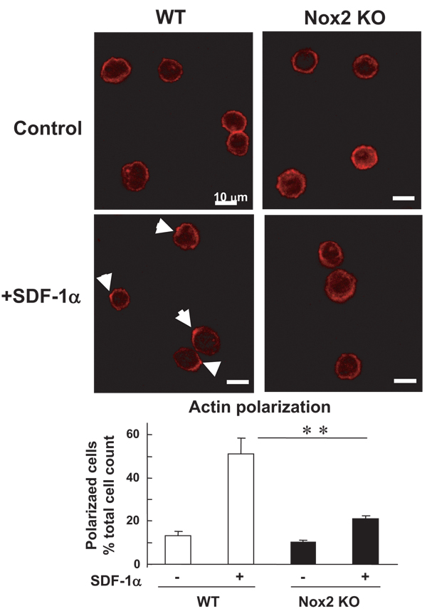
Figure 7.
SDF-1α–induced F-actin polarization is impaired in Nox2−/− BM stem/progenitor cells in vitro. Upper panel, freshly isolated WT- and Nox2−/− BM c-kit+Lin− cells in suspension were stimulated with or without SDF-1α for 5 minutes, fixed, and then stained for Alexa Fluor 568 conjugated phalloidin, which is visualized by confocal microscopy. Arrows indicate the polarized F-actin. Original magnification is ×630 and bars show 10 µm. Lower panel, quantitative analysis of SDF-induced F-actin polarization in WT- and Nox2−/− BM c-kit+Lin− cells. Data are expressed as percentage of the total 100 cells from 4 randomly selected fields (×100; n=4, **P<0.01).

