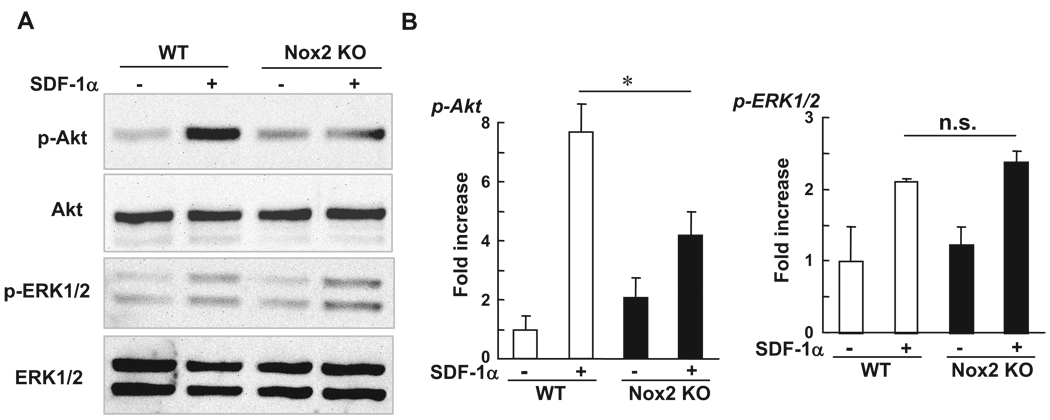
Figure 8.
Phosphorylation of Akt induced by SDF-1 is inhibited in Nox2−/− BMCs. Freshly isolated WT- and Nox2−/− BMCs were serum depleted for 4 hours and then stimulated with SDF-1α (300 ng/mL) for 5 minutes. Lysates were used for Western analysis with antiphospho-Akt (Ser473), phospho-ERK1/2, total Akt, or total ERK1/2 antibodies. Data are expressed as fold increase over the unstimulated WT cells (n=3, *P<0.05).

