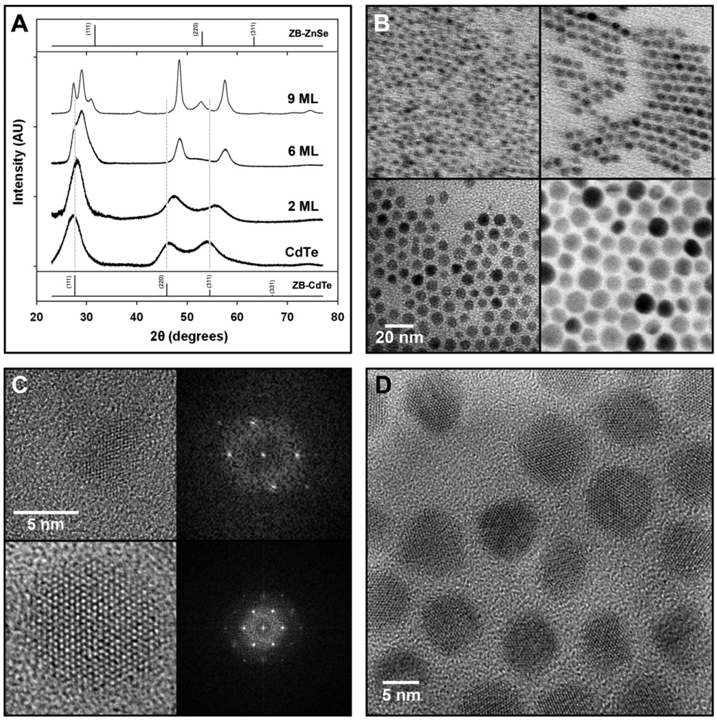
Figure 4. Lattice structures of strain-tunable QDs as determined by power x-ray diffraction and high-resolution transmission electron microscopy.
(A) Powder x-ray diffraction patterns for 3.8 nm CdTe QDs, and (CdTe)ZnSe QDs with 2, 6, or 9 monolayers of shell growth, from bottom to top. Bulk diffraction peaks for zinc blende (ZB) CdTe and ZnSe are indexed at the bottom and top, respectively, and vertical lines correspond to the major diffraction lines of CdTe. The x-ray wavelength was 1.78897 Å. (B) Transmission electron micrographs of 3.8 nm CdTe cores capped with 0 (top left), 2 (top right), 6 (bottom left), and 9 (bottom right) monolayers of ZnSe. (C) High-resolution transmission electron micrographs of 3.8 nm CdTe QDs (top) and (CdTe)ZnSe QDs with 6 monolayers of shell (bottom). Fast-Fourier transform spectra of these materials are shown on the right. (D) HRTEM of (CdTe)ZnSe QDs with 6 monolayers of shell, showing all QDs to be oriented with the (001) lattice plane parallel to the substrate. The absorption and emission spectra of these (core)shell QDS are provided in Supplementary Figure 4, and their simulated diffraction and particle size distribution data are shown in Supplementary Figures 4 and 5. The results obtained from a line-wdith analysis of the diffraction peaks are summarized in Supplementary Table 1.

