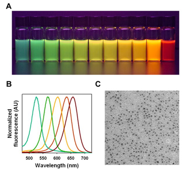
Figure 2.
Fluorescence emission and electron-microscopy structural properties of CdTe core QDs prepared by using multidentate polymer ligands in a one-pot procedure. (A) Color photograph of a series of monodispersed CdTe QDs, showing bright fluorescence from green to red (515 nm to 655 nm) upon illumination with a UV lamp. (B) Normalized fluorescence emission spectra of CdTe QDs with 35–50 nm full width at half maximum (FWHM). (C) Transmission electron micrograph of QDs showing uniform, nearly spherical particles.

