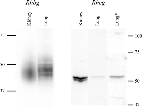
Fig. 2.
Immunoblot of kidney and lung tissue for Rhbg and Rhcg. Membrane-enriched fraction of cellular lysate from mouse lung and mouse kidney was examined by immunoblot analysis for expression of Rhbg (left) and Rhcg (right). On the left, 1.25 μg of kidney and 40 μg of lung protein were used. Lung Rhbg expression was ∼3% of renal Rhbg expression after adjustment for amount of protein loaded. On the right, 20 μg of lung tissue and 25 μg of kidney protein were used. The lanes labeled kidney and lung are from the same immunoblot and are reproduced with original intensity. The lane labeled “lung*” uses contrast-enhancement of the lung lane to more clearly show the Rhcg band. Lung Rhcg expression was ∼6% of renal Rhcg expression after adjustment for amount of protein loaded.

