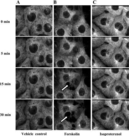
Fig. 2.
Forskolin stimulation of sACI/II increases a cytosolic cAMP pool that reorganizes microtubules sufficient to disrupt PMVEC barrier function. PMVECs that are stably expressing GFP-α-tubulin were infected with an adenovirus expressing the sACI/II. Live cell confocal imaging was performed for 35 min in control cells and in forskolin (10 μmol/l) or isoproterenol (1 μmol/l) treated cells. A: in vehicle control cells, microtubules were not reorganized, and endothelial barrier was not disrupted (n = 4). B: in forskolin-treated cells, peripheral microtubule networks were reorganized, and endothelial cell gap formation (arrows denote endothelial barrier disruption as a consequence of reorganization of peripheral microtubule networks, n = 9). C: in isoproterenol-treated cells, microtubules were not reorganized, and the endothelial barrier was not disrupted (n = 5). The scale bars represent 25 μm.

