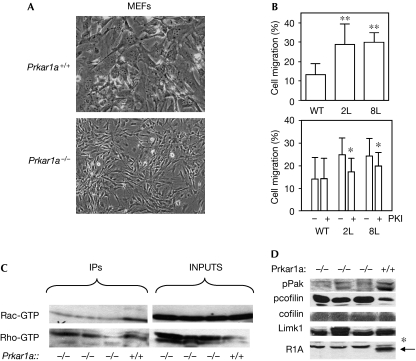
Figure 1.
Loss of Prkar1a causes altered cell morphology and enhanced migration independent of Rho GTPases. (A) Phase-contrast images of Prkar1a+/+ and Prkar1a−/− MEFs illustrating altered cell morphology induced by the loss of Prkar1a. (B) Top: migration of WT MEFs and two Prkar1a−/− MEF lines (2L and 8L) in a Boyden chamber assay. **P<0.01 compared with WT cells. Bottom: PKA inhibitor (PKI) reduces migration of knockout but not of WT MEFs. *P<0.05 for drug-treated versus vehicle-treated cells. (C) Measurement of total and activated (GTP-bound) Rac and Rho. (D) Immunoblot analysis of phospho-Pak (pPak), pcofilin, total cofilin, Limk1 and Prkar1a (R1A) levels in knockout (−/−) and WT (+/+) lysates. Note the presence of a nonspecific band (*) in the Prkar1a lane. IPs, immunoprecipitates (of activated proteins); Limk1, LIM domain kinase 1; MEF, mouse embryonic fibroblast; PKA, protein kinase A; WT, wild type.

