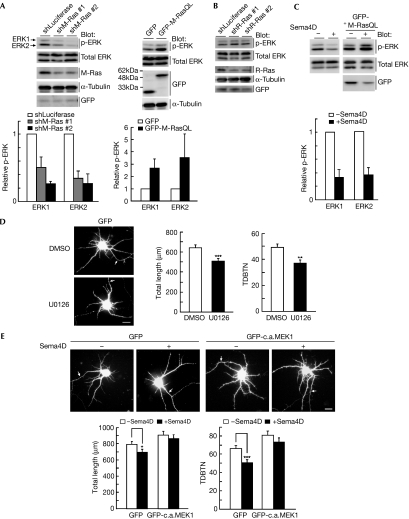
Figure 3.
Sema4D reduces dendrite growth through the down-regulation of extracellular signal-regulated kinase activity. (A) Cortical neurons were transfected with control shRNA (shLuciferase), M-Ras shRNA (#1 or #2), green fluorescent protein (GFP) or GFP-fused M-Ras(Q71L), before plating, using nucleofection technology; cell lysates at 7 days in vitro (DIV) were analysed by immunoblotting with antibodies against phospho-ERK, ERK, M-Ras, α-tubulin and GFP. (B) Cortical neurons were transfected with control shRNA or R-Ras shRNA (#1 or #2) with GFP, before plating, using nucleofection technology; cell lysates at 7 DIV were analysed by immunoblotting with the indicated antibodies. (C) Cortical neurons transfected with or without M-Ras(Q71L) were treated at 7 DIV with or without Sema4D for 1.5 h; cell lysates were analysed by immunoblotting with antibodies against phospho-ERK and ERK. (D) Cortical neurons transfected with GFP at 5 DIV were treated with or without 20 μM MAPK/extracellular signal-regulated kinase kinase (MEK) inhibitor U0126 for 2 d and fixed at 7 DIV. (E) Cortical neurons were transfected with GFP or GFP-fused constitutively active MEK1 (GFP-c.a.MEK1) at 5 DIV. Neurons at 7 DIV were stimulated with Sema4D for 1.5 h and fixed. Transfected cells are shown by GFP fluorescence. Arrows indicate axons; scale bar, 20 μm. Total length of dendrites and total dendritic branch tip number (TDBTN) were measured. The results are the means±s.e.m. of three independent experiments (n=40, *P<0.05, **P<0.01 and ***P<0.005). ERK, extracellular signal-regulated kinase; shRNA, short hairpin RNA.

