
Figure 4. Cytokine secretion in response to poly(I:C) stimulation is significantly increased in SDR DCs.
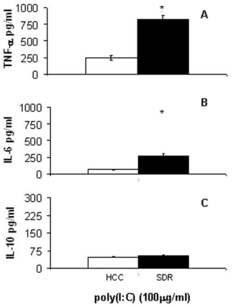
CD11c+ cells from HCC (□) or SDR (■) mice were stimulated in culture with 100μg/mL Poly(I:C). At 18h post culture supernatants were analyzed by cytometric bead array. Data are presented as the amount of TNF-α (A), IL-6 (B) and IL-10 (C) in the supernatants of HCC or SDR DC cultures. Asterisks (*) indicate that the difference between groups is statistically significant (p < 0.01) in repeated experiments. Results are representative of three experiments. (n= 12 mice per group, per experiment).