
Figure 6. NGF and NT-3 Stimulate Tyrosine Phosphorylation of the trk Proto-Oncogene Product.
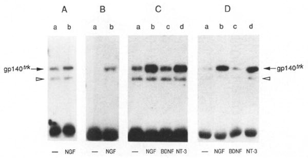
Quiescent E25-48 cells were incubated for 10 min at 37°C with DMEM alone (lanes a) or with DMEM containing 50 ng/ml NGF (lanes b), BDNF (lanes c), or NT-3 (lanes d). Cells were lysed in RIPAE buffer containing 0.5 mM sodium orthovanadate, immunoprecipitated with a rabbit polyclonal antibody elicited against a peptide corresponding to the carboxy-terminal domain of gp140trk (Martin-Zanca et al., 1989) and analyzed by 8% SDS–polyacrylamide gel electrophoresis. The electrophoresed samples were transferred to nitrocellulose filters and blotted with anti-trk antiserum (A) or anti-phosphotyrosine monoclonal antibody 4G10 (UBI) (B–D) as described in Experimental Procedures. (C) depicts tyrosine phosphorylation of gp140trk after stimulation with baculovirus-expressed and BDNF and NT-3, (D) after stimulation with COS cell–expressed BNDF and NT-3. The resulting nitrocellulose filters were exposed to Kodak X-Omat film for 8 hr at −70°C with the help of an intensifying screen.