
Figure 6. Analysis of p75NTR and Pax-2 Expression at E13.5 and E15.5.
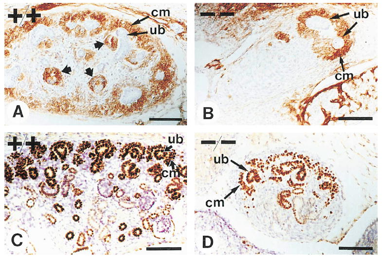
(A and B) Sections from E13.5 wild-type (A) and mutant (B) animals were stained with antibodies to the p75NTR receptor. Expression was apparent in wild-type and mutant animals in condensing mesenchymal cells (cm) surrounding the ureter branches (ub). In wild-type animals, p75NTR expression was also observed in renal vesicles (A, wide arrows). (C and D) Sections from E15.5 wild-type (C) and mutant (D) animals were stained with antibodies to Pax-2, which is expressed both in the ureteric epithelium and upon induction in the condensing mesenchyme. Only a small part of a section from the wild-type kidney is shown, whereas the whole rudiment of an α8-deficient animal is visible. Note that in mutant animals the ureter had formed very few branches but Pax-2 expression was induced. Scale bars, 50 μm.