
Figure 3. Sensory Populations.
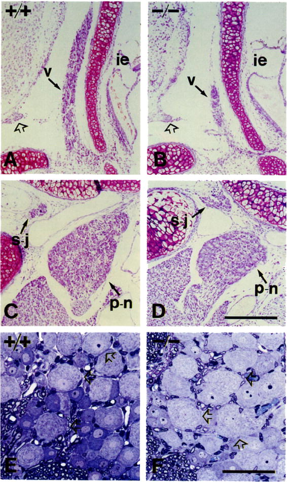
(A and B) Vestibular ganglia (v) of P0 +/+ (A) and −/− (B) mice. Symbols: ie, inner ear; open arrow, acoustic nerve.
(C and D) Superior–jugular (s–j) and petrosal–nodose (p–n) ganglia of +/+ and −/− mice. The pair petrosal–nodose (placode derived) is reduced in volume in mutant animals. The intracranial pair superior–jugular (neural crest derived) appears unaffected.
(E and F) PI5 mice L5 dorsal root ganglia sectioned in araldite and stained with toluidine blue. Note the presence of large lightly stained neurons in the mutant (open arrows), consistent with the presence of normal numbers of muscle spindles in the thigh skeletal muscles.
Scale bars, (A–D) 200 μm; (E and F) 50 μm.