
Figure 5. NPY Expression.
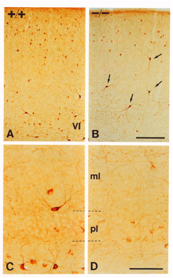
(A and B) Cerebral cortex stained with NPY antibodies of +/+ (A) and −/− (B) littermates. Note that there is some reduction in numbers of intermediate to small neurons expressing NPY in the mutant animal. However, the large neurons expressing high levels of NPY (arrows) persist.
(C and D) CA1 region of the hippocampus of P17 wild-type (C) and mutant (D) animals. The number of immunoreactive interneurons is reduced in all areas of the hippocampal formation Note that cells in mutant animals express very low levels of peptide. Abbreviations: pl, pyramidal cell layer; ml, molecular layer.
Scale bars, (A and B) 200 μm; (C and D) 50 μm.