Figure 4.
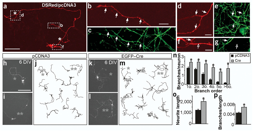
DsRed and tau-1 are colocalized and branch numbers are increased in hippocampal cells in the absence of FAK at 6 DIV in vitro. At early stages, most of the neurites in these cultures express tau-1 in neurons transfected with control (shown here) and Cre- or FRNK-expressing plasmids (not shown). (a,b,d,f) Expression of DsRed and pCDNA. (c,e,g) Tau-1 immunoreactivity. (b,d,f) Higher magnification images corresponding to the boxed regions indicated in a. Most branching neurites at these stages are developing axons as they express the axon-specific marker tau-1 (∼95%, n = 10 neurons). Arrows help to position the colocalization. Filled arrows indicate axons (tau-positive neurites), open arrows indicate dendrites (tau-negative neurites), white asterisks indicate branchpoints. (h–p) fakflox deletion in mouse hippocampal cultures. (h–m) Fluorescence images and representative drawings of neurons transfected with control pCDNA3 (h–j) or Cre plasmid (k–m) at 6 DIV. (n) Quantification of branches at increasing branch orders per neuron. *P < 0.01 to P < 0.0001 versus control by two-tailed t-test (n = 92 neurons from four cultures in n–p). (o) Quantification of the total neurite length per neuron expressed in µm.*P < 0.0005 versus control (control 1,241.36 ± 93.24, mutant 2,018.04 ± 169.44). (p) Quantification of the total branches per µm of axon. *P < 0.0005 versus control (control 0.0045 ± 0.0003, mutant 0.0067 ± 0.0005). Note that, as described above, most branching neurites at these stages are developing axons (∼95%), although a few of them could be dendrites. Data are the mean ± s.e.m. Scale bars, 200 µm (n,m); 100 µm (a); 10 µm (b–g).

