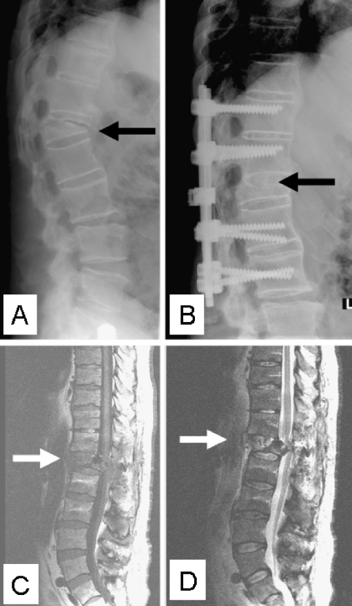
Fig. 3.
Lateral plain radiograph with a kyphotic discovertebral AL at the thoracolumbar junction in a 60-year-old male AS patient (a). Radiograph after posterior instrumentation and fusion of the symptomatic AL is also shown (b). T1- (c) and T2-weighted (d) sagittal MR images accurately reveal involvement of the surrounding structures. The lesion has resulted in a severely narrowed spinal canal, with dural compression, that clinically resulted in a postoperative partial peroneal nerve palsy of his right leg

