
Figure 3.
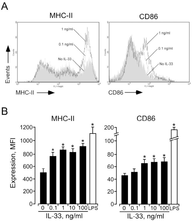
IL-33-activated DCs show increased expression of MHC II and CD86. A, After 24 h culture of DCs with medium (shaded area) or IL-33 (0.1 ng/ml (broken line) and 1.0 ng/ml (solid line)), MHC II and CD86 expression levels were examined by flow cytometry. B, After 24 h culture of DCs with IL-33 or LPS (1 μg/ml), MHC II and CD86 expression levels were examined. *p<0.05, compared to medium alone, n=5.