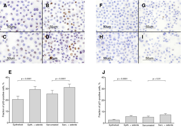
Figure 2.
Nuclear translocation of p53 and p21. A-E: Immunocytochemical analysis of p53 performed on cytospin samples. A: Epithelioid cells without selenite. B: Epithelioid cells treated with 10 μM selenite for 24 h. C: Sarcomatoid cells without selenite. D: Sarcomatoid cells treated with 10 μM selenite for 24 h. E: Fraction of cells with p53-positive nuclei after 24 h, as assessed by two independent observers. Bars show the 95% confidence interval. χ2-tests were employed. F-J: Immunocytochemical analysis of p21 performed on cytospin samples, as an additional readout for p53 activity. F: Epithelioid cells without selenite. G: Epithelioid cells treated with 10 μM selenite for 24 h. H: Sarcomatoid cells without selenite. I: Sarcomatoid cells treated with 10 μM selenite for 24 h. J: Fraction of cells with p21-positive nuclei after 24 h, as assessed by three independent observers. Bars show the 95% confidence interval. χ2-tests were employed. Three independent experiments were performed.

