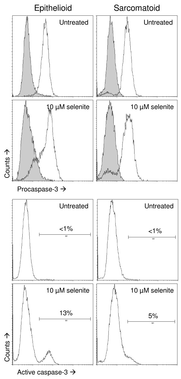
Figure 6.
Caspase-3 activation as determined by flow cytometry. Top four panels: flow cytometric analyses of procaspase-3. Sarcomatoid and epithelioid cells showed a similar baseline expression. In both cell types, a subpopulation lost expression after selenite treatment. Gray histograms show the negative controls for the immunostaining. Bottom four panels: flow cytometric analyses of caspase-3 activation. Selenite treatment caused the appearance of a distinctly positive subpopulation in the epithelioid cells, whereas the sarcomatoid cells showed a small positive subpopulation that was not distinctly separated from the main peak. Three independent experiments were performed. All eight panels are derived from the same experiment.

