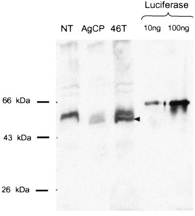
Figure 5.
Western blot analysis of luciferase protein expression. Guts from the nontransformed recipient mosquito line (NT), the AgCP line, or an AeCP line (46T) were dissected from mosquitoes 24 h after a blood meal. Gut homogenates were analyzed by Western blotting with an antiluciferase antibody. The two lanes on the right contain recombinant luciferase (10 and 100 ng), used as a positive control. Migration of molecular weight markers is indicated on the left. The arrowhead indicates the position of migration of the predicted truncated protein (see text), detected only in the transgenic lines.

