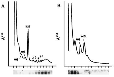
Figure 6.
Association of the luciferase mRNA with polysomes. Guts from mosquitoes transformed with the AeCP construct were dissected at 24 h after a blood meal, homogenized, and divided into two equal aliquots. One was analyzed on a sucrose gradient without further additions (A), and the other was analyzed after dissociation of polysomes by addition of EDTA to 10 mM (B). The upper part of each panel shows the A254 profile of the gradient after centrifugation, and the lower part shows the Northern blot analysis of each fraction of the gradient by using a radioactive luciferase probe. The short vertical marks below the abscissae indicate the position of each fraction. The numbers above the absorption profiles indicate the position of migration of the ribosomal subunits (40S and 60S), whole ribosomes (80S), or the number of ribosomes per polysome.

