
Figure 2.
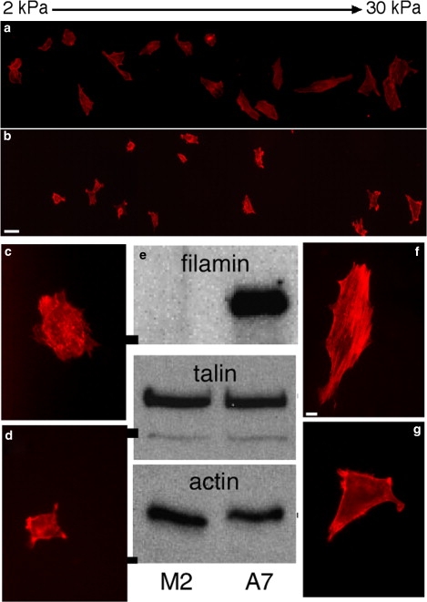
Morphological change of melanoma cells on stiffness gradient gels. Shapes of A7 (a) and M2 (b) cells bound to a PA gradient gel coated with collagen I and fibronectin, taken 24 h after plating. Gel stiffness increases from left to right. Scale bar is 40 μm. The full width of the gel is 1.8 mm, and its stiffness ranges from 1 to 30 kPa. Higher magnification images show examples of A7 (c and f) and M2 (d and g) cells on soft (c and d, 0.5 kPa) and stiff (f and g, 15 kPa) gels. Scale bar for these images is 10 μm Western blot (e) shows equal levels of talin and actin expression in M2 and A7 cells, but no filamin A expression in M2 cells. Molecular mass bars on left of image are 200 kDa for filamin and talin, and 40 kDa for actin.