
Figure 1.
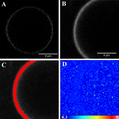
GUV containing 99% POPC and 1% BODIPY TMR-labeled PI(4,5)P2. (A) Confocal microscopy image of a GUV in the absence of profilin. (B) Intensity map of the GUV membrane. (C) The highlighted pixels correspond to a molecular brightness of 1.070. (D) Brightness map of the same GUV membrane.