
Figure 7.
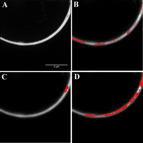
Brightness analysis of GUV made of 92% POPC and 8% PI(4,5)P2. The concentration of BODIPY TMR labeled PI(4,5)P2 was 1% molar ratio. (A) Fluorescence intensity map of the GUV. (B) The pixels highlighted correspond to the molecular brightness of monomers (1.071). (C) The highlighted pixels have an intermediate brightness of 1.376 and correspond to clusters in the membrane. (D) The highlighted pixels have the highest brightness (2.263) and correspond to areas of larger clusters.