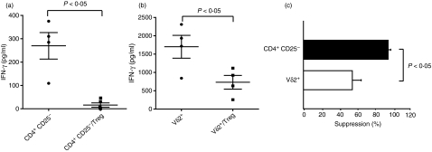
Figure 4.
CD4+ CD25high T cells suppress antigen-specific CD4+ CD25− and Vδ2+ T-cell responses. (a) Fluorescence-activated cell sorting (FACS) sorted CD4+ CD25− (2.5 × 10⁴ cells) isolated from tuberculin-skin-test-positive (TST+) donors were stimulated with purified protein derivatve (PPD; 10 μg/ml) in the presence or absence of CD4+ CD25high T cells (Tregs) at a 1 : 1 effector : suppressor ratio. Interferon-γ (IFN-γ) in 5-day culture supernatants was determined by enzyme-linked immunosorbent assay (ELISA). Mean values ± SEM of four independent experiments are shown. (b) FACS-sorted Vδ2+ T cells (2.5 × 10⁴ cells) isolated from TST+ donors were stimulated with BrHPP (10 μm) plus interleukin-2 (25 U/ml) and PPD (10 μg/ml) in the presence or absence of CD4+ CD25high T cells (Tregs) at 1 : 1 effector : suppressor ratio. IFN-γ in 5-day culture supernatants was determined by ELISA. Mean values ± SEM of four independent experiments are shown. (c) Percentage suppression was determined as follows: (IFN-γ in cultures with CD4+ CD25high)/(IFN-γ in cultures without CD4+ CD25high) × 100. Means ± SEM are shown for four independent experiments.

