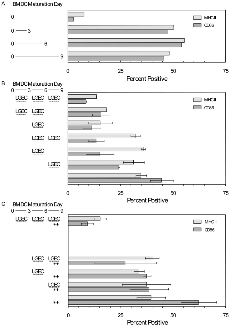
Figure 3. Ex vivo maturation of dendritic cells from rat bone marrow monocytes and influences of soluble factors from rat lacrimal epithelial cells.
A. DC were matured from rat bone marrow monocytes ex vivo for 3, 6, or 9 days. Surface expression of MHC Class II and CD86 was determined by flow cytometry. B. DC were matured from rat bone marrow monocytes for 9 days. Microporous inserts containing ratLGEC were present during the indicated intervals. Values presented are means ± ranges for two replicate cell preparations. C. DC were matured as in panel B. Symbols indicate that LPS was present at a concentration of 10 µg/mL during day 8 and day 9. Values presented are means ± ranges for two replicate cell preparations.

