Figure 8.
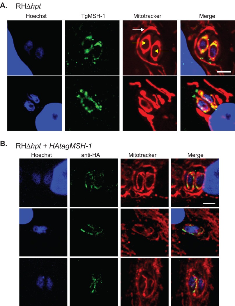
Mitochondrial localization of TgMSH-1. The mitochondria of intracellular wild-type (RHΔhpt) parasites and of the HFF host cells were stained with 300 nM MitoTracker Red CMX-ROS and the nuclei were stained with Hoechst Stain. A. TgMSH-1 was labeled with serum generated against amino acids 1752–2095 of TgMSH-1 and was visualized with an alexa fluor-488 conjugated goat anti-mouse antibody. Yellow arrows point at the 2 intracellular parasites and the white arrow indicates the host mitochondria surrounding the parasitophorous vacuole, which contains the parasites. Signal from each fluorophore and a merged image is shown. Scale bar, 4 microns. B. HA-tagged TgMSH-1 was visualized in parasites expressing the tagged protein using an anti-HA primary antibody in combination with an alexa fluor-488 conjugated goat anti-rabbit secondary antibody. Scale bar, 4 microns.

