Figure 1.
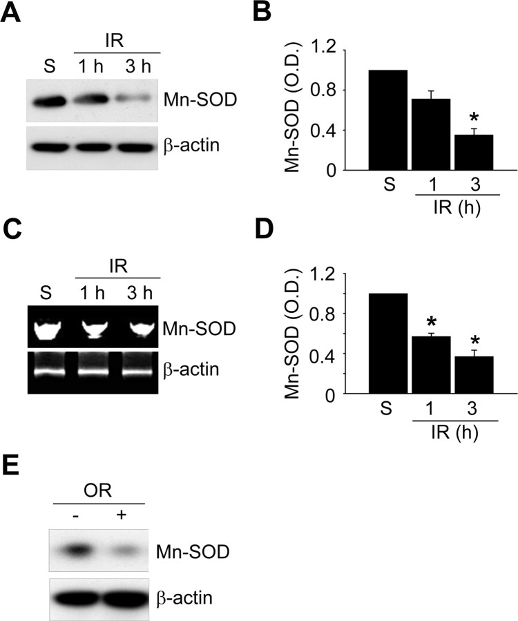
Mn-SOD expression is significantly reduced by ischemic reperfusion in a mouse MCAO model. A, Western blot analysis of the protein level of Mn-SOD in mouse brains after 1 and 3 h of reperfusion following MCAO with an antibody against anti-Mn-SOD antibody. Whole-cell protein extracts were obtained from the cerebral cortex and caudate–putamen (except the hippocampus). B, Summary graph depicting the change in the Mn-SOD protein level relative to β-actin. *p < 0.05 (n = 4 per group). C, The mRNA level of Mn-SOD in mouse brains after 1 and 3 h of reperfusion following MCAO was determined by RT-PCR using a specific primer against the mouse Mn-SOD gene. Total RNA was prepared from the cerebral cortex and caudate–putamen (except the hippocampus), and equal RNA loading was confirmed using mouse β-actin-specific primers. D, Summary graph depicting the change in Mn-SOD mRNA levels relative to β-actin. *p < 0.05 (n = 4 per group). E, Western blot analysis of the protein level of Mn-SOD in primary cortical neurons subjected to reoxygenation 3 h after OGD for 2.5 h. IR, Ischemic reperfusion; S, sham; O.D., optical density; OR, oxygen–glucose deprivation/reoxygenation; C, control.

