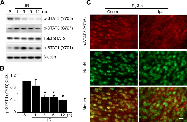
Figure 2.
STAT3 is significantly downregulated by ischemic reperfusion in a mouse MCAO model. Whole-cell protein extracts were obtained from the cerebral cortex and caudate–putamen (except the hippocampus) of male mouse brains after 1, 3, 6, and 12 h of reperfusion following MCAO. A Western blot was performed with an antibody against phospho-STAT3 (Y705), phospho-STAT3 (Ser-727), phospho-STAT1 (Y701), and total STAT3. A, Representative phospho-STAT3 (Y705), phospho-STAT3 (Ser-727), phospho-STAT1 (Y701), and total STAT3 blots and a β-actin blot showing equal protein loading. B, Summary graph depicting the change in phospho-STAT3 relative to total protein loading. *p < 0.05 (n = 4 per group). C, Phosphorylation status of STAT3 (red) and exhibition of the neuronal marker NeuN (green) from ischemic regions (cortex) on the contralateral and ipsilateral sides shown by immunohistochemistry analysis. Scale bar, 20 μm. IR, Ischemic reperfusion; S, sham; O.D., optical density; NeuN, neuron-specific nuclear protein.

