
Figure 1.
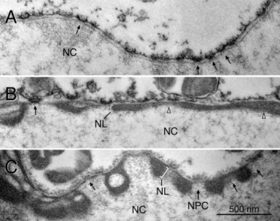
TEM of the NEs of lamin A-expressing oocytes. Prelamin A was expressed in oocytes by nuclear injection of plasmid. TEM sections of isolated oocyte nuclei are shown. The lamina (NL) (brackets in B and C) forms a thick electron-dense layer in oocytes expressing lamin A (B and C), which leaves the NPCs (arrows) free. The lamina in noninjected control oocytes is hardly discernible (A). NC: nuclear content; arrows point to NPCs; open triangles in B point to adjacent lamina layers of different thickness. Images were taken at the same magnification.