
Figure 2.
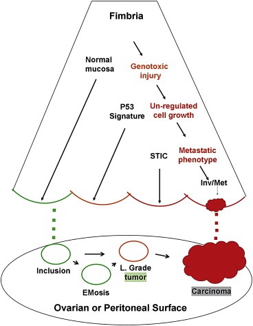
– Schematic of the distal fallopian tube and two pathways to pelvic serous cancer. In one pathway, passive transfer of normal cells from the fimbria (green dots) leads to ovarian cortical inclusions that are usually quiescent but may ultimately evolve into serous malignancy via endometriosis or low‐grade tumors. In the second pathway, several tubal mucosal epithelial entities, including normal secretory cells (green), p53 signatures (yellow), and tubal intraepithelial carcinoma (STIC, red) are linked by a series of connected biologic events including unrepaired genotoxic injury, unregulated cell growth, and eventually, acquisition of a metastatic phenotype. The latter permits spread to the ovarian or peritoneal surfaces (red dots).